


See every cell
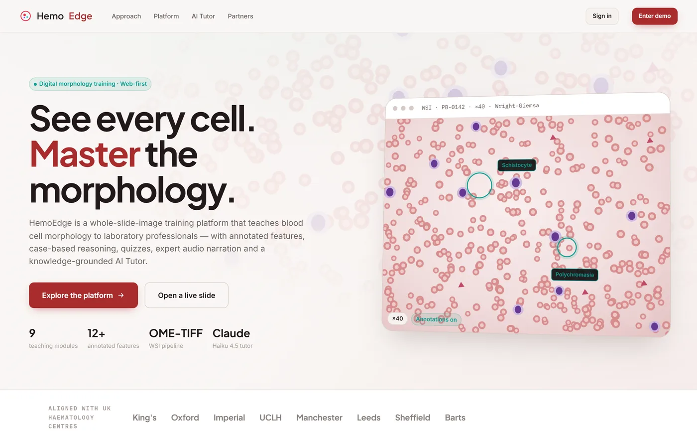
An image-first platform for teaching blood cell morphology in the browser: deep-zoom whole slide images, expert annotations, case-based reasoning, quizzes and an AI tutor that cites its own sources.
Sector
- Healthcare education
- Digital pathology
Platform
- Learner application
- Educator console
What we did
- Product modelling
- UX + UI design
- Front-end engineering
- Grounded AI design
Curriculum
- 9 teaching modules
- 12+ annotated features
What is the platform for?
Laboratory professionals learn to recognise abnormal blood cells down a microscope. HemoEdge moves that training into a browser without losing the thing that makes it work — looking closely at a real film, with an expert pointing.
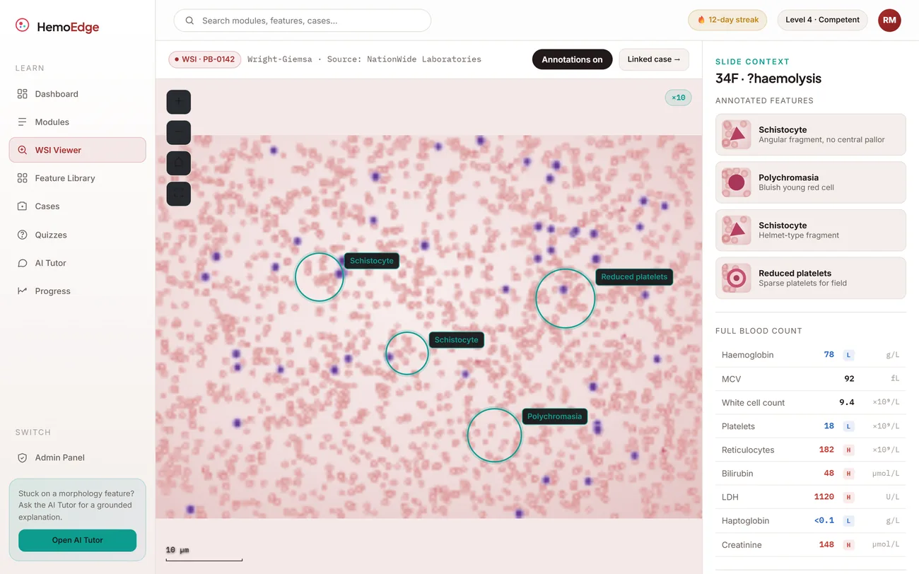
A microscope, a curriculum and a tutor in one screen
The slide is the centre of the product. Everything else - the annotations, the clinical context, the blood count, the tutor - is arranged around the image rather than in a separate lesson somewhere else.
Why is morphology training so hard to scale?
Three gaps show up in every lab. Together they mean two trainees in two hospitals can finish the same grade having seen entirely different material, with no way to compare what either of them can actually do.
| Training activity | At the microscope | Modelled in HemoEdge |
|---|---|---|
| Access to teaching slides | Whatever is on site this week | A shared library, revisitable at any time |
| Expert commentary | Whoever is free to sit with you | Annotations and audio attached to the slide |
| Curriculum | Learned opportunistically | Nine structured modules, foundation to advanced |
| Clinical context | Remembered, or looked up separately | Case history and blood count beside the film |
| Answering a question | Ask a colleague, if one is nearby | AI tutor answering from reviewed teaching notes |
| Assessment | A conversation, rarely recorded | Image-based quizzes with tracked competency |
What can you actually click?
The platform covers the whole learner journey and the educator's side of it.
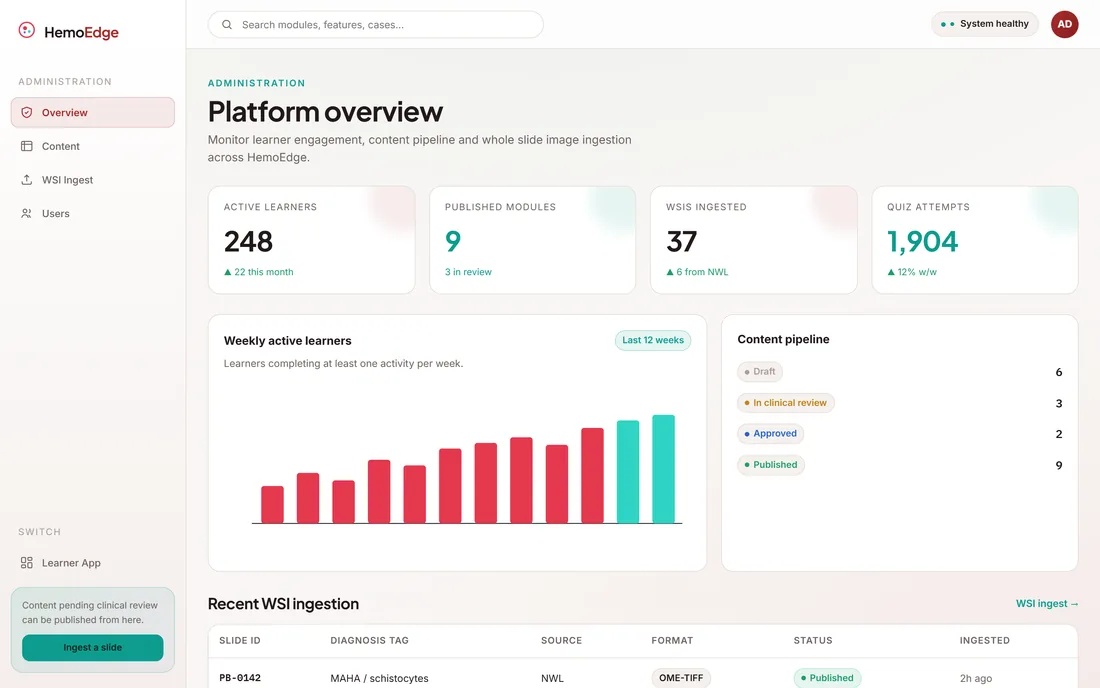
Whole slide imaging
OME-TIFFDeep-zoom peripheral blood films delivered in an open, metadata-rich format suited to digital microscopy, annotation and viewing in a browser.
- Deep zoom
- Scale bar
- Annotations toggle
Feature library
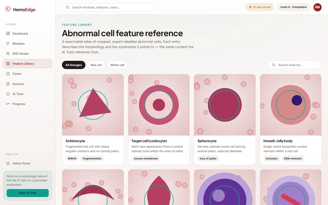
12+ featuresCropped, labelled images of key abnormal cells with morphology notes and the syndromes they point to.
- Named features
- Syndrome notes
Structured modules
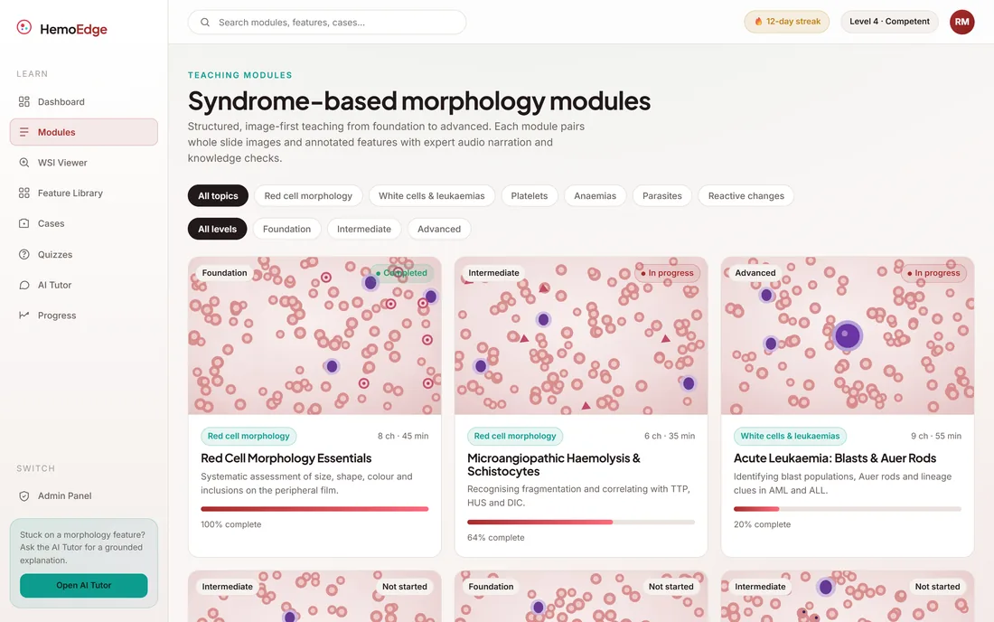
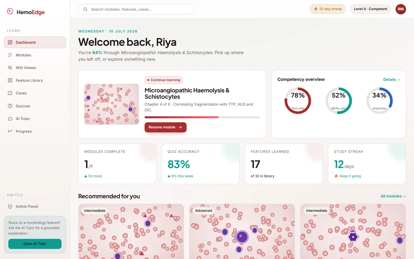
9 modulesA graded curriculum from foundation to advanced, so learning follows a route instead of whatever came through the lab.
- Foundation to advanced
- Chaptered
Case-based learning
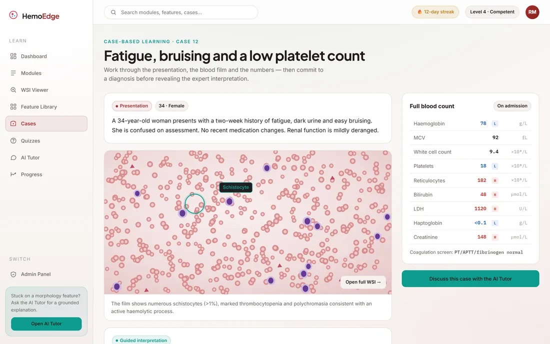
ReasoningClinical scenarios that pair a film with the patient history and lab values, then work through the reasoning the way an experienced reader would.
- Blood count
- Patient context
- Guided reasoning
Quizzes & assessment
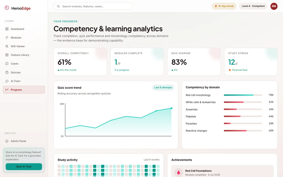
MeasuredImage-based questions with instant feedback and explanations, building a competency record rather than a completion tick.
- Instant feedback
- Competency tracking
Grounded AI tutor
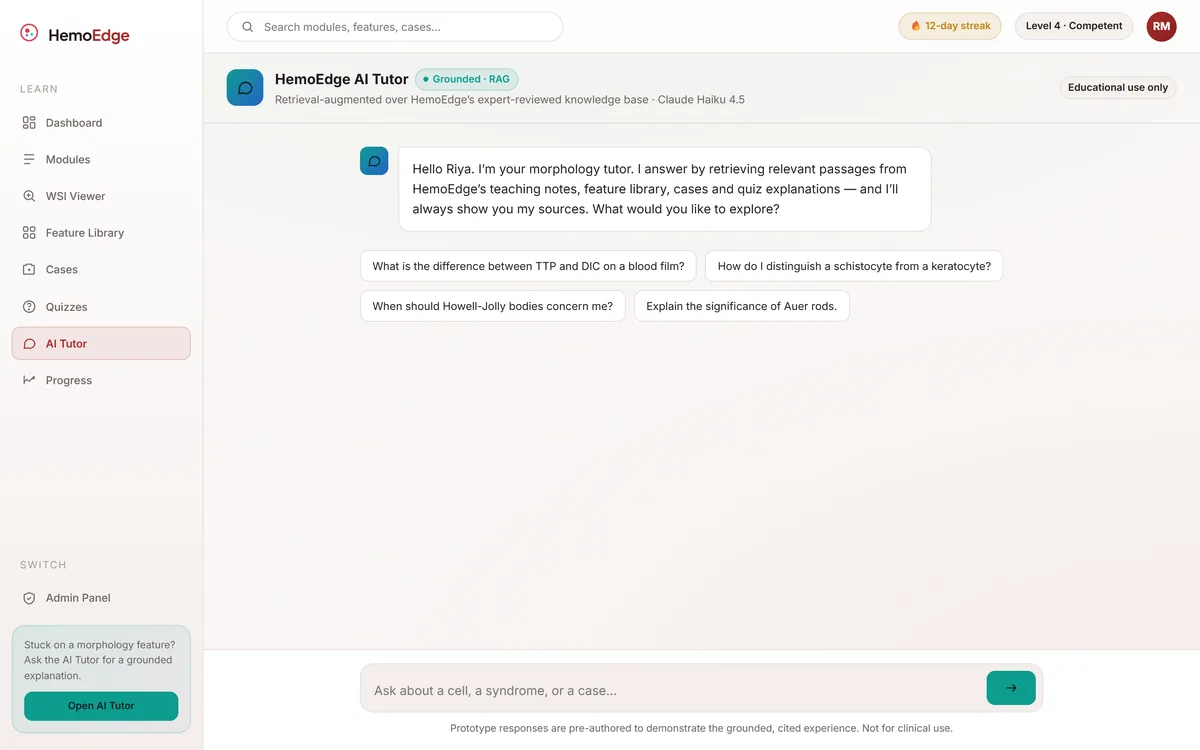
CitedA retrieval-augmented assistant answering from expert-reviewed material, showing the source behind every answer instead of improvising.
- Knowledge base
- Shown sources
- Audio narration
How does a slide become measured competence?
One pipeline, from a physical film on a bench to a competency a training lead can evidence.
What does an expert annotation actually add?
The same field of a peripheral blood film, with and without the teaching layer. Drag the handle: on one side it is a picture of some cells, on the other it is a lesson pointing at the four that matter.
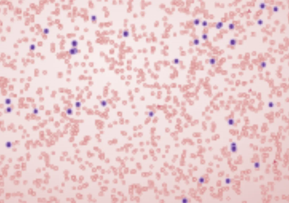
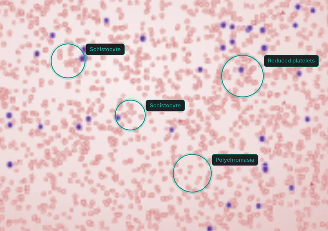
The annotation layer is authored by haematologists and toggles on and off over any slide in the library.
What does any clinical training platform need?
Six things decide whether clinical staff trust a training tool enough to use it. They came out of modelling this one, and they apply to any platform teaching a visual, judgement-heavy skill.
- The artefact at full fidelity. If the learner cannot zoom to the magnification they work at, they are studying a picture of the skill rather than the skill.
- Expert attention made explicit. An annotation that points at the cell that matters is the whole value a supervisor adds. Ship that, not a caption.
- Context beside the image, not behind a tab. Morphology is read against the history and the blood count; separating them teaches the wrong habit.
- AI that cites, or none at all. In a clinical setting an unsourced answer is worse than no answer. Ground it in reviewed material and show the source.
- Assessment that produces a record. Competency has to be demonstrable across cohorts and sites, which means it must be measured, not asserted.
- Provenance on every teaching claim. A learner has to know which consultant reviewed a feature and when — content nobody will put their name to does not get trusted, and in a lab that means it does not get used.
What does the interface look like, and why?
The palette is taken from the subject itself - a deep stain crimson for the brand and anything to do with the blood film, a clinical teal reserved for progress, correctness and the tutor. Everything else is warm neutral, so the pinks in the slide image are always the most saturated thing on screen.
Aa
Primary typeface
Plus Jakarta Sans
A B C D E F G H I J K L M N O P Q R S T U V W X Y Z
a b c d e f g h i j k l m n o p q r s t u v w x y z
0 1 2 3 4 5 6 7 8 9 & ? ! £ $ €
34pxSee every cellH124pxSection headingH220pxModule and card headingH316pxLead copy and teaching notesLead14pxBody text, feature notes and table cellsBody12pxSLIDE CONTEXT · ANNOTATED FEATURESEyebrowColour and type are read from the running platform rather than sampled off a screenshot. Lab values are set with tabular figures so a full blood count reads as a column, and the high and low flags stay the only coloured thing in the table. Inter and Plus Jakarta Sans are open-licence typefaces; the specimen above falls back to the nearest available face if they are not installed on your device.
What is it built on?
The pieces that make deep-zoom slides and a grounded tutor work in a browser.
Building a clinical training platform
What laboratories and training providers ask us first.
Ask us yoursHow long does it take to build a digital training platform?
A first working version usually takes eight to sixteen weeks, depending on how much of the content pipeline is in scope. The application itself is rarely the long pole - digitising source material, and getting expert reviewers through enough of it to make the curriculum useful, is what actually sets the date.
How much does a platform like this cost?
It depends on scope. The biggest drivers are whether image conversion and hosting are included, how many learner and educator roles exist, and whether assessment has to satisfy an external standard. Shanti Infosoft scopes it on a short call and returns a fixed quote within 48 hours.
How do you stop an AI tutor from making things up?
By not letting it answer from general knowledge. The tutor is retrieval-augmented: it answers from expert-reviewed teaching notes, feature entries and case context, and shows the source under each answer. In a clinical setting an unsourced answer is worse than no answer, so grounding and visible citation are design requirements rather than features.
Can whole slide images really work in a browser?
Yes, with the right format. Slides are converted to a tiled, metadata-rich format so the browser only fetches the tiles for the current view and magnification. That is what makes pan-and-zoom over a very large scan feel immediate without downloading the whole image.
Related work
Have training that only works in person?
Tell us how your business actually works and we will tell you honestly what it takes to build. See more of our work or what we do.
Book a 15-minute call